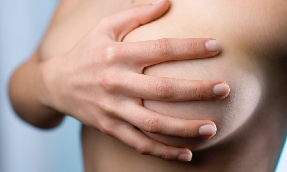
One in eight women in the United States will receive a breast cancer diagnosis. The risk factors for breast cancer include older age; genetics; menstrual, pregnancy, and menopause history; alcohol use; and hormone replacement therapy.
The earlier breast cancer is discovered, the higher the likelihood it can be cured. When caught early, the five-year survival rate can be as high as 90%. The most important factor in breast cancer outcome is whether the cancer has spread to the lymph nodes, so catching the cancer early has a major impact on survival.
For this reason, women have been urged to have mammograms to screen for breast cancer. Whether this should start at age 40 (American College of Obstetricians and Gynecologists), age 40-45 (American Cancer Society), or age 50 (US Preventive Health Services Task Force) remains controversial.The cancers detected by the ultrasound were more likely to be invasive and were more frequently node negative than those detected by mammography.
The answer may be ultrasonography.
Ultrasounds, often used when a woman is pregnant to check the status of her baby, may be a useful, low-cost alternative to mammography, according to a recent study.
Over 2600 women were recruited from 20 sites. Each woman underwent three rounds of mammography and of ultrasonic screening exams. These were done at the start of the study and then repeated 12 and 24 months later. The results were ranked on a scale from “normal exam” to “highly suggestive of malignancy.” Women were followed up appropriately, depending on their results.
During the study, 110 women were diagnosed with breast cancers. Mammography and ultrasound each detected the same numbers of cancers, and there were no significant differences between the two screening methods with respect to breast cancers detected in women with dense breasts, or in women of different age groups.
The authors suggest that ultrasound is an alternative well worth considering, especially in developing countries where women do not have access to mammography and therefore have no screening other than physical exam available to them. Ultrasound equipment is significantly less expensive than mammography and has the advantage of being portable.
Ultrasounds may also be a useful supplemental test for women with dense breast tissue who do not meet criteria for MRI studies (recommended for women at high risk with dense breast tissue) or who cannot tolerate MRI.
One barrier to implementation to an ultrasound-based screening program, according to the authors, is the rate of false positives but they maintain that the rate of false positives will fall when the results of earlier ultrasounds are available for comparison.
The authors hope that their findings will be used to address the health needs of women in developing countries, giving them the benefits of early detection and potential for a complete cure.