Dr. Barasch is the Director of the Cardiac Echo Laboratories at the Hermann Heart Center and the University of Texas Medical School, Houston.
Echocardiography is a modern technique that allows the physician to evaluate the heart without inserting any tubes or wires. This "non-invasive" method, like the sonogram pregnant mothers now routinely receive, uses sound waves, which reflect off the heart structures (hence the term echo) and are recorded to produce an image of the heart.
Computer processing of the precise arrival times of the sound signals provides a two-dimensional view of a "slice" through the heart. In the twenty years since it was first introduced, echocardiography has evolved into many specialized formats: color Doppler, three dimensional, tissue imaging and volumetric echocardiography.
Echocardiography can be very helpful in the diagnosis of:
Significant Heart Murmurs
First and foremost, doctors use echocardiography to evaluate the seriousness of heart murmurs. Murmurs are extra sounds, heard by the doctor through the stethoscope, that are produced as blood flows through an opening changed by disease or birth defect. Echocardiography records these blood flow measurements and converts them into pressure gradients, the difference in pressure between one side of the opening (at a valve, for example) and the other side, thus telling the doctor how severe the damage is. With the help of this test, the physician can determine how leaky or narrowed the valve is and identify which patients might benefit from medicines or corrective surgery.
Heart Enlargement
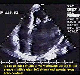
For patients with enlarged hearts, the echocardiogram will accurately measure the degree of enlargement of the upper collecting chambers (atria) or the lower pumping chambers (ventricles). If you have an atrial septal defect (ASD), a hole in the wall separating the two upper collecting chambers, transesophageal echocardiography (which uses a probe mounted on a flexible tube that is inserted through the mouth into the esophagus located just behind the heart) will identify the type of defect, its size, its direction and possible associated abnormalities. Similarly, for ventricular septal defect (a hole between the pumping chambers), which can be congenital or acquired (usually after a heart attack), characteristics of the defect can be determined.
Heart Infections and Emboli
Echocardiography is so sensitive that it can also detect mild murmurs not otherwise heard. This early warning system can alert your doctor to future problems. Infections of the heart (infective endocarditis) often occur at the site of a defective heart valve and, until echocardiography, it was difficult to pinpoint the infection's location. Artificial valves, for example, are prone to infection and echocardiography enables the doctor to better monitor their status. Sometimes, one type of echocardiography, transthoracic echocardiography (TTE), using a probe positioned on the chest, will be normal and transesophageal electrocardiography (TEE) will be needed to detect the problem.
Echocardiography has also been helpful in detecting cases where the outside lining of the heart is infected (pericarditis).
For a patient with an embolus [any undissolved material (usually a blood clot) carried by the blood to other organs, such as the brain in stroke patients], echocardiography can be used to identify from where in the heart or the aorta the embolus originated.
Heart Failure
In congestive heart failure, the heart is unable to pump efficiently and effectively. Echocardiography helps the doctor determine the cause of the problem. Are the upper chambers out of synch with the lower ones? And at what part of the heart's cycle? Are the heart chambers enlarged? Are the walls of the heart moving poorly? Echocardiography provides important information for the management of these problems.5
Coronary Artery Disease (CAD)
Three coronary arteries supply the heart muscle with oxygen and nutrients. When these arteries are blocked by fat-laden plaques, the resultant coronary artery disease (CAD) can produce chest pain or an acute heart attack [acute myocardial infarction (AMI)], both of which can be diagnosed by echocardiography.6 In patients who are having a heart attack, a resting echocardiogram can identify the location, extent and consequences of the AMI on the mechanical functioning of the heart.
If combined with stress testing, where the doctor asks you to exercise prior to the test, echocardiography can indicate the functional status of the heart more accurately than the standard exercise electrocardiogram stress test. Post-treadmill echocardiography or echo following pharmacologic stress with dobutamine, a drug which raises heart blood flow, are the most utilized protocols for exercise and pharmacological stress, respectively, in the United States. They are more likely to detect patients with less severe cases of coronary artery disease than the standard ECG stress test. Moreover, the dobutamine stress test can also indicate which areas of the heart muscle are alive and well. This information gives the doctor the ability to predict the post-operative success of bypass surgery.
Fainting Spells
Rest echocardiography and/or stress echocardiography are valuable tools which can help the doctor determine whether your heart can handle the stress of surgery.
Other Uses
Adult patients who suffer from diseases such as congenital heart disease, lung disease, connective tissue disorders (lupus) need to be followed indefinitely for heart problems and echocardiography is the best way to monitor them. Recently, patients on certain medicines found to have heart consequences, for example the diet medicine, phen-fen, were screened with echo to see if they had sustained heart damage. Donor hearts for heart transplant patients can also be evaluated with Doppler echocardiography.
Conclusion
A careful history and physical examination by a doctor is the foundation of a cardiac evaluation but echocardiography is becoming an increasingly valuable technique that can help the physician decide whether a particular medical or surgical therapeutic option is in the patient's best interest.